Intermittent fasting shows promise in managing obesity and cardiovascular health
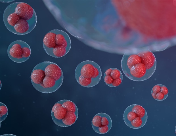
A team of scientists led by the University of Granada (UGR), the Public University of Navarra (UPNA) and the CIBER has s...

A team of scientists led by the University of Granada (UGR), the Public University of Navarra (UPNA) and the CIBER has s...

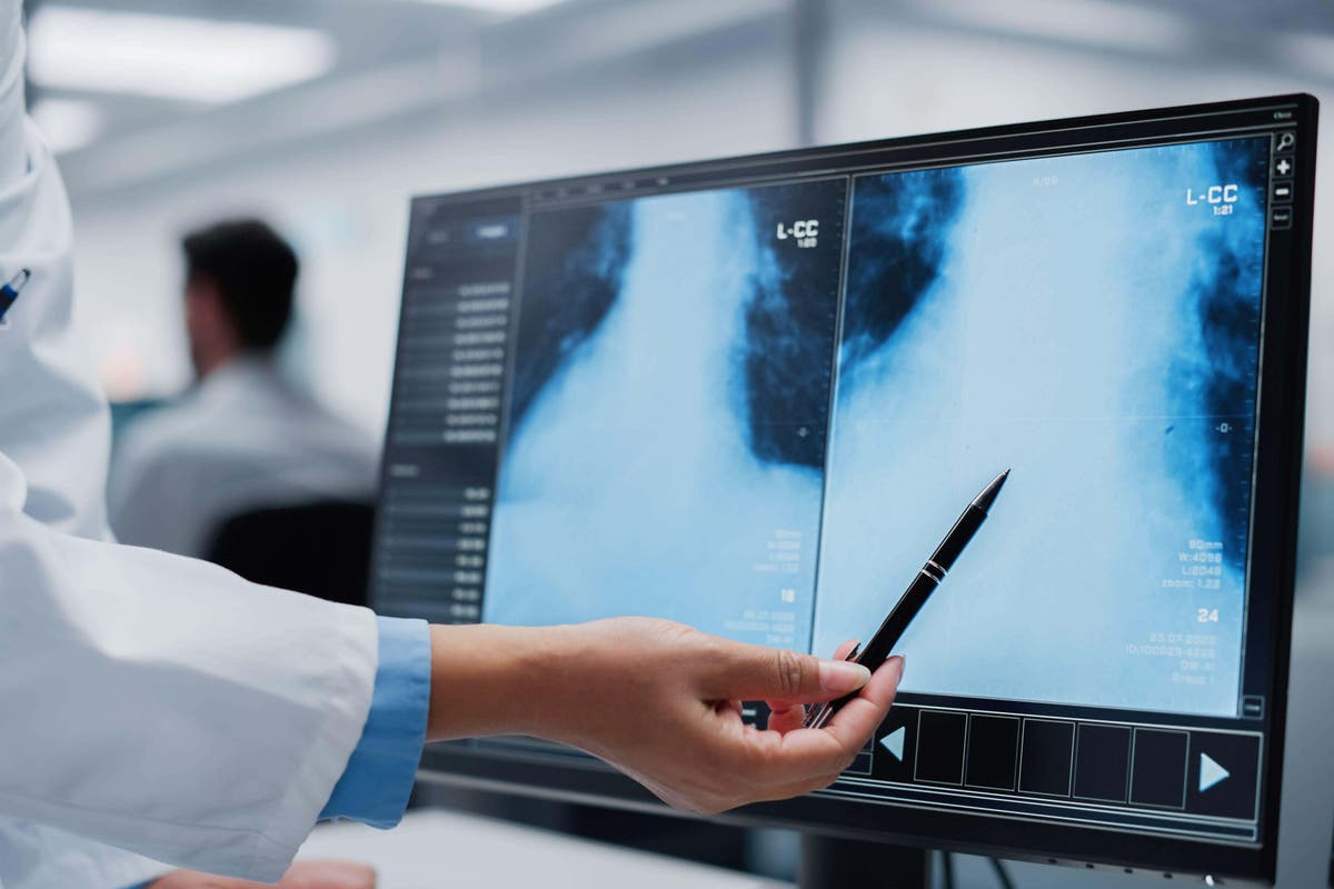
AI-supported mammography screening significantly increases breast cancer detection rates while maintaining comparable re...


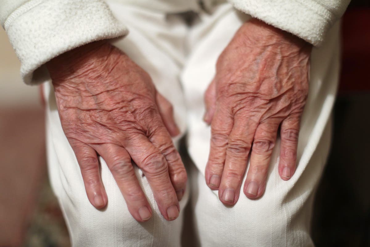
Time-restricted eating (TRE) is safe and feasible for weight management but offers no additional benefits over a Mediter...

Steve Sosnick, Interactive Brokers' Chief Strategist tells Squawk Box Asia the massive rally in quantum computing stocks...

What Are The Health Risks Associated With Wildfire Smoke?...
Cancer remains a significant public health challenge, with the GLOBOCAN 2020 report estimating a staggering 19.3 million...

The melding of visual information (microscopic and X-ray images, CT and MRI scans, for example) with text (exam notes, c...

A common vitamin might be silently affecting your health. Uncover the startling revelations from recent studies that cou...
The discovery of a new type of stem cell in the brain could usher in better treatments for the deadliest brain tumor....
Photoimmunotherapy is an innovative cancer treatment that combines phototherapy with immunotherapy to selectively target...

Jim Caron of Morgan Stanley Investment Management explains what's behind the steepening yield curve and what it's tellin...

The fires burning across Los Angeles have sent billowing plumes of black smoke into the air, posing a major health threa...
The Centers for Disease Control and Prevention (CDC) has selected University of California San Diego as one of three par...

Using a new artificial intelligence method, researchers at Columbia University Vagelos College of Physicians and Surgeon...

Local health and aged care lawyer and advocate Catherine Henry looks ahead to 2025 reforms...

Electrical stimulation of the spinal cord is a promising strategy for reestablishing walking after spinal cord injury, r...
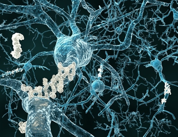
The gene neuropilin2 encodes a receptor involved in cell-cell interactions in the brain and plays a key role in regulati...
A new study has shown that sleep deprivation can inhibit the brain's ability to suppress unwanted memories and intrusive...

A University of California, Irvine-led research team has discovered intricate molecular mechanisms driving the RNA proce...
Scientists at La Jolla Institute for Immunology (LJI) have discovered how a mutated gene kicks off a dangerous chain of ...

Facing high employee turnover and an aging population, nursing homes have increasingly turned to robots to complete a va...

Julian Emanuel of Evercore ISI says Trump's economic policies will be moderated and influenced by the bond markets. His ...
A new study led by Winship Cancer Institute of Emory University and Abramson Cancer Center of University of Pennsylvania...
Clinicians have successfully used custom-made 3D printed bone scaffolds, printed on-site at The University of Queensland...

"Mad Money" host and former hedge fund manager, Jim Cramer, provides stock traders with all manner of investing advice....

'Mad Money' host Jim Cramer weighs in on stock including: Pfizer, Vertex, Serve Robotics, Quantumscape, and Powell Indus...
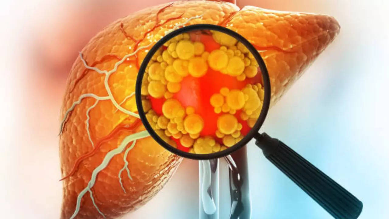
Could an ordinary carrot improve the treatment of type 2 diabetes? A new study from SDU suggests so....

'Mad Money' host Jim Cramer looks at the ticking clock for TikTok as it faces down its sale deadline....
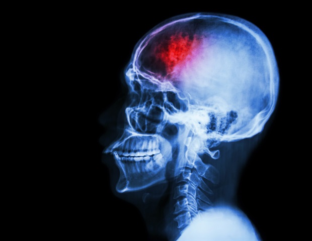
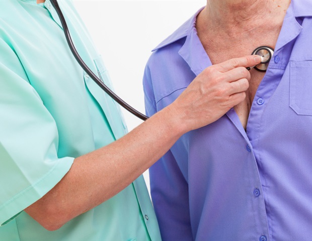
Older people wait an average of just over a month to see a neurologist for specialty care after being referred by their ...
Childhood stress can impact the epigenetic profile of sperm. These results may also have practical implications for futu...

Olive leaf extract may reduce blood pressure, improve blood lipids and help the way our bodies handle glucose. But the e...

'Mad Money' host Jim Cramer looks at today's market action....
In a rare case, a recipient of a donated organ died of cancer after contracting the illness from the donor. Doctors said...
Women are less likely to receive a lung transplant and spend an average of six weeks longer on the waiting list, accordi...

Prostate Cancer UK says ministers have a 'moral imperative' to change the rules so GPs can proactively speak to at risk ...

Taimur Baig of DBS Group Research discusses the challenges facing the Chinese economy. He says Beijing's stimulus measur...

US longshoremen reached a tentative contract agreement with ports and shippers Wednesday, averting a strike that could h...

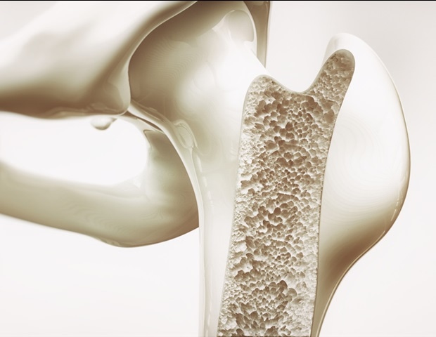
A new study by researchers at the UNC Thurston Arthritis Research Center at the UNC School of Medicine has identified 13...
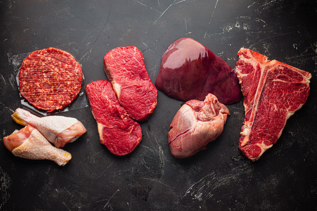
Learn which supplements might be necessary and how adding dairy or organ meats can help meet nutritional needs....
Samsung Biologics to offer ADC services at new dedicated facility Extended collaboration reflects successful partnership...

Capitol Police arrested a man in line for the viewing of Jimmy Carter in the Capitol Rotunda on Wednesday who had a mach...

Massive wildfires burning in the Los Angeles area have filled the air with a thick cloud of smoke and ash, prompting air...

The high school seen in movies including Carrie was badly damaged in the Los Angeles wildfires, but early reports that P...

Two workers at a state-run hospital in West Virginia have been charged in connection with the death of a 61-year-old non...

'Mad Money' host Jim Cramer looks at today's market action....

The S&P 500 eked out a narrow gain on Wednesday. The NYSE is closed on Thursday in honor of late former President Jimmy ...
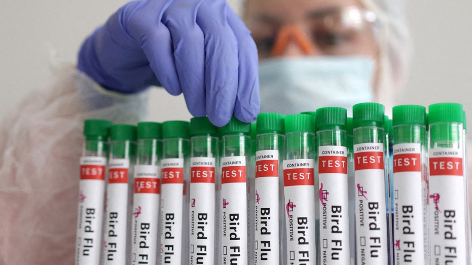
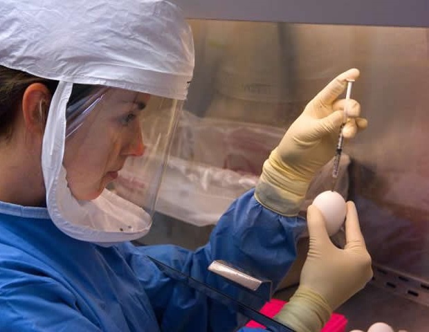
Texas officials on Wednesday confirmed several dead birds that were discovered earlier this month near Austin tested pos...

"Fast Money" is America's post-market show. Hosted by Melissa Lee and a roundtable of top traders, "Fast Money" breaks t...

'Mad Money' host Jim Cramer looks at the ticking clock for TikTok as it faces down its sales deadline....
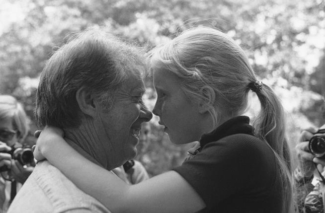
Amy Carter was 9 when she joined her parents in walking in Jimmy Carter's inaugural parade in 1977. She then lived in th...

A Hollywood A-lister has come to the help of an immigrant living in Minnesota as he fights a serious illness....
Roughly 13% of people move to a nursing home in the year after diagnosis...
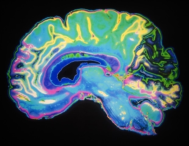
Medics used advanced MRI scans to identify different areas of tumours....

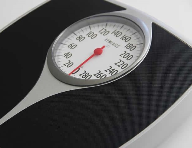
'Aerobic fitness is more important for mortality risk than body weight.'...

From facials to scalp treatments, Zen Luxury Scalp & Hair Spa has created a haven for anyone looking to unwind....
:upscale()/2025/01/08/012/n/1922729/tmp_UDBLJD_742ee19303d7ef2a_GettyImages-1303468858.jpg)
Gym equipment wipes: You use them, we use them, and we all hope the person who used the machine before us used them, too...

COLUMBUS, Ohio (AP) — Ohio Gov....

"This new ability will impact every industry, from biomedicine to finance,'' Dr. Alex Zhavoronkov said....

Registered nurse Delilah Clyburn-Hill and certified nursing assistant Kylah Beard have been charged with felony neglect ...

An outbreak of the virus in northern China has prompted some online alarm but experts say the risk of another Covid-like...