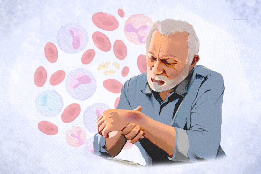
Leukemia is a general term for cancers affecting blood cells. The specific type of leukemia is determined by the type of blood cell involved and the rate at which the cancer progresses. Leukemia is most commonly found in adults over 55, but it is also the most prevalent cancer in young people under 15.
Blood stem cells develop into either lymphoid or myeloid stem cells. Lymphoid stem cells produce lymphocytes, a type of white blood cell that fights infections, while myeloid stem cells produce red blood cells, platelets, granulocytes, and monocytes, with the latter two also being white blood cells. Before maturing into blood cells, lymphoid and myeloid stem cells first differentiate into blast cells, such as lymphoblasts and myeloblasts, which are immature blood cells.
Lung Cancer: Symptoms, Causes, Treatments, and Natural Approaches Liver Cancer: Symptoms, Causes, Treatments, and Natural Approaches In leukemia, abnormal blast cells are overproduced and do not mature into functional blood cells. These leukemia cells often grow and survive more effectively than normal cells, gradually crowding out and suppressing the production of healthy blood cells. The progression and impact on normal cells vary depending on the type of leukemia.
1. Acute Myeloid Leukemia (AML) 2. Chronic Myeloid Leukemia (CML) 3.
Acute Lymphoblastic Leukemia (ALL) ALL is the most common leukemia in children, making up about 80 percent of cases in this age group. Most children with ALL have the B-cell subtype. 4.
Chronic Lymphocytic Leukemia (CLL) Anemia: Anemia (insufficient red blood cells) may present as fatigue, weakness, pallor (pale skin), malaise, dyspnea on exertion (shortness of breath with activity), tachycardia (rapid heartbeat), and chest pain during exertion. Thrombocytopenia: Thrombocytopenia (insufficient platelets) can lead to bleeding from mucous membranes, easy bruising, purpura (small red or purple spots on the skin), nosebleeds, bleeding gums, and heavy menstrual periods. Spontaneous bleeding, including in the brain or abdomen, may also occur.
Granulocytopenia or neutropenia: Neutropenia (insufficient neutrophils, a type of white blood cells) increases the risk of infections from bacteria, fungi, and viruses. Symptoms may include fevers and severe or repeated infections. The exact cause of the fever is often unclear, but granulocytopenia (insufficiency in a type of white blood cell) can lead to rapidly worsening and serious bacterial infections.
Pancytopenia: Pancytopenia is characterized by low levels of all three above types of blood cells. Swelling of the lymph nodes, liver, or spleen: When leukemic cells infiltrate organs, it can enlarge the liver, spleen, and lymph nodes. Fever and chills.
Unexplained weight loss. Night sweats. Loss of appetite.
Joint pain. Bone pain. ALL is staged according to leukemia cell maturity and the type of lymphocyte involved.
AML is staged using the French-American-British (FAB) system, which accounts for how many leukemia cells there are and what size, number of healthy cells, chromosome changes, and other genetic issues. CLL is staged with the Rai system, which considers the blood’s lymphocyte count, the degree of spleen, lymph node, and liver enlargement, and whether there is anemia or low platelet count. CML is staged according to how many diseased cells are in the blood and bone marrow.
Age: Age is a risk factor that differs among types. The highest incidence rates for ALL occur in children and adolescents under 15 years old, and children to middle-aged people are more at risk for CML. However, adults 50 years or older are more at risk for AML and CLL.
Sex: Men are more at risk for all four types of leukemia than women. Genetic syndromes: These include Down syndrome, Fanconi anemia, Bloom syndrome, ataxia-telangiectasia, neurofibromatosis type 1, Shwachman-Diamond syndrome, congenital dyskeratosis, Kostmann syndrome, Wiskott-Aldrich syndrome, Li-Fraumeni syndrome, Klinefelter syndrome. Genetics and family history: Inherited genetic mutations present from birth may increase the risk of developing AML.
Race: In the United States, ALL is more common among whites and Hispanics, and CLL is more common among whites than other races. Gene alterations: Most DNA changes associated with AML develop during a person’s lifetime rather than being inherited at birth. Changes in specific genes such as FLT3, c-KIT, and RAS are common in AML cells.
These changes can prevent bone marrow cells from maturing properly or cause them to grow uncontrollably. Smoking: Smoking is associated with about a 50 percent increased risk of leukemia, possibly due to the presence of the carcinogen benzene in tobacco. Radiation exposure or chemotherapy: People who have received radiation therapy or chemotherapy (e.
g., with drugs called etoposide and alkylating agents) have a higher risk of leading to AML, known as treatment-related or therapy-related AML. People exposed to very high levels of radiation, such as survivors of atomic bomb blasts or nuclear reactor accidents, are also at increased risk of AML and CML.
Leukemia from radiation exposure takes many years to develop, and most people treated with radiation for cancer do not develop the disease. CML has also been reported in those who have undergone excessive diagnostic X-rays or CT scans. Benzene exposure: Benzene is present in unleaded gasoline and is used in the chemical industry.
People can be exposed to benzene through their work, the environment, or by using certain products. It is found in paint strippers, adhesives, forest fire smoke, and volcanic gas. Blood and bone marrow disorders: People with certain blood disorders, such as myelodysplastic syndrome and myeloproliferative disorders, and bone marrow diseases are at higher risk for developing AML and CLL.
HTLV-1 infection: Infection with the human T-cell leukemia/lymphoma virus, type 1 (HTLV-1) raises the risk of developing T-cell ALL, a rare form of ALL. Epstein-Barr virus (EBV): EBV is associated with a type of ALL. In the United States, EBV is the virus that commonly causes infectious mononucleosis.
Weakened immune system due to immunosuppressive drugs. Exposure to herbicides: Researchers have identified a possible link between CLL and exposure to herbicides such as Agent Orange used during the Vietnam War. The U.
S. Department of Veterans Affairs provides disability compensation to those exposed to this herbicide. Chromosome changes: In most cases of CLL, changes are found in chromosomes, commonly involving deletions such as the loss of part of chromosome 13.
Other affected chromosomes can include 11 and 17, and occasionally there is an extra chromosome 12 (trisomy 12). Blood Tests Complete blood count (CBC): A complete blood count measures the number and quality of white blood cells, red blood cells, and platelets. The presence of immature blood cells (blasts) in the blood, which are not normally seen, may lead doctors to suspect leukemia.
Blood chemistry tests: Blood chemistry tests measure specific chemicals in the blood to help identify liver or kidney issues caused by leukemia and help stage the disease. In leukemia, levels of certain chemicals, such as blood urea nitrogen, creatinine, phosphate, alanine aminotransferase, and uric acid, may be elevated. Blood clotting tests: Blood clotting tests evaluate how well the body can clot blood, as abnormal levels of clotting factors can be associated with leukemia.
Key tests include fibrinogen level, prothrombin time (PT), partial thromboplastin time (PTT), and international normalized ratio (INR). Kidney function panels. Liver function panels.
Peripheral blood smear: Blood cells are stained and examined under a light microscope to reveal their number, size, shape, and type, as well as the pattern of white blood cells and the ratio of blast cells to maturing and mature white blood cells. Quantitative immunoglobulin test: The quantitative immunoglobulin test measures the concentration of antibodies (immunoglobulins) in the blood, which are produced by B-cells to help protect the body from infections. Other Tests Bone marrow tests: Bone marrow testing typically involves two steps that are usually done simultaneously.
These samples are usually taken from the hip bone and examined to detect chromosomal and cellular changes. First, during bone marrow aspiration, a liquid sample is collected. Next, a bone marrow biopsy removes a small bone piece with marrow.
Immunophenotyping (flow cytometry): Immunophenotyping is a technique that uses fluorescent labels to sort and classify cells. This method allows doctors to quickly examine many antibodies on thousands of cells in a single sample, and it’s useful for identifying unique features of leukemia cells. Imaging tests: Doctors use imaging tests such as X-rays, computed tomography (CT) scans, magnetic resonance imaging (MRI), or ultrasounds to rule out other causes of symptoms and to check for leukemia cell masses in the chest that could affect breathing or blood circulation.
Spinal tap: A spinal tap (lumbar puncture) involves using a needle to collect cerebrospinal fluid, which is then examined for cancer cells. Cytogenetic and Molecular Studies Cytogenetic karyotyping: Cytogenetic karyotyping identifies chromosomal abnormalities, helping doctors confirm a leukemia diagnosis and determine its type or subtype. These results also assist in planning the appropriate treatment.
Fluorescence in situ hybridization (FISH): FISH uses fluorescent DNA probes to detect chromosomal abnormalities and genetic changes in leukemia cells. It helps differentiate between leukemias with similar appearances but distinct genetic profiles. Polymerase chain reaction (PCR): PCR is a very sensitive test that can find a single leukemia cell among 100,000 cells.
It amplifies specific gene segments to detect DNA mutations, inversions, or deletions associated with certain leukemia types. It aids in diagnosing leukemia and assessing its prognosis. Quantitative polymerase chain reaction (qPCR): The qPCR test is a highly sensitive method used to find and measure the amount of a specific gene, BCR::ABL1 (related to the Philadelphia chromosome), in blood or bone marrow samples.
It can detect even tiny amounts of this gene, which may not be visible with other tests. Biomarker testing: Biomarker testing or molecular/genomic testing analyzes the DNA or RNA sequence in cancer cells to identify genetic changes. This information helps assess risk, predict prognosis, and guide treatment decisions, including the need for intensive or novel therapies.
DNA sequencing: DNA sequencing tests blood or marrow samples for mutations in certain genes. Identifying mutations or their absence helps assess the patient’s prognosis. Beta-2 microglobulin test (B2M): Beta-2 microglobulin is a small protein produced by various cells and can be measured through a blood chemistry test.
Disseminated intravascular coagulation (DIC): DIC is a leukemia complication where blood clotting proteins malfunction, causing both clotting and bleeding. It is commonly linked to acute promyelocytic leukemia but can occur with other leukemia types. Regular lab monitoring and fibrinogen replacement with cryoprecipitate are crucial for patient survival.
Severe infections: Immunosuppression due to chemotherapy, stem cell transplantation, or the leukemia alone raises the risk of severe infections. Other cancers: Leukemia survivors have an increased risk of developing subsequent cancers. Research has found a 5.
6 percent risk of any cancer occurring within 30 years after leukemia. The most frequent second cancers in childhood leukemia survivors are other types of leukemia or lymphoma. Organ dysfunction: Leukemia cells can invade organs.
Chemotherapy: Chemotherapy uses anti-cancer drugs, either injected or taken orally, to target fast-growing cells, affecting both cancerous and healthy cells. It’s the primary treatment for many types of leukemia. The typical treatment for AML is known as the “7+3” regimen, where patients receive cytarabine continuously for seven days and an anthracycline drug (either daunorubicin or idarubicin) for three days.
Targeted therapy: In recent years, new drugs have been developed that specifically target certain parts of cancer cells. These targeted drugs work differently from traditional chemotherapy and usually have different side effects. Imatinib, nilotinib, dasatinib, bosutinib, and ponatinib are tyrosine kinase inhibitors (TKIs) that block the abnormal protein produced by the Philadelphia chromosome.
Most patients respond well to TKIs, which are the standard treatment for CML, so acute chemotherapy is usually only necessary if the disease progresses to an accelerated phase or blast crisis. These drugs have also been successful in combination with chemotherapy for treating the blast phase, which previously had a poor prognosis. Stem cell transplant: For some patients in remission who can handle intensive chemotherapy, the doctor might suggest a stem cell transplant during the consolidation phase.
Interferon therapy: Interferons are natural substances produced by the immune system, and interferon-alpha is a manmade version that mimics this natural substance. Interferon therapy helps slow the growth of leukemia cells. Active surveillance: CLL progresses more slowly than other types of leukemia and usually doesn’t significantly shorten a person’s lifespan.
Early treatment isn’t helpful unless the patient meets specific criteria for starting therapy. Radiation therapy: Radiation therapy uses high-energy radiation to destroy cancer cells. It is used to treat a person with CLL with an enlarged lymph node, spleen, or other organ.
A positive mindset can help patients handle the emotional challenges of leukemia, potentially leading to better overall well-being. It can also encourage patients to stick with their treatment plans and follow medical advice. Medicinal Herbs Feverfew (Tanacetum parthenium): Feverfew, also known as bachelor’s button , is a daisy-like plant commonly found throughout North America.
Malignant stem cells play a key role in the development and persistence of AML. The compound parthenolide , derived from feverfew, has been found to selectively trigger the death of these AML stem cells, unlike standard chemotherapy, which often misses these vital cells and leaves the root cause of leukemia unaddressed. However, parthenolide’s effectiveness in medical treatments was limited as the body didn’t absorb it well.
In 2019, researchers at Birmingham University developed a new parthenolide derivative that is better absorbed by the body and has promising effects against leukemia based on in vitro and in vivo studies. So far, clinical trials haven’t been completed on this derivative’s effectiveness in treating leukemia. Korean red ginseng: A 2009 study found that treating U937 human leukemia cells with Korean red ginseng extract slowed their growth and triggered cell apoptosis (programmed cell death).
A 2016 study tested a water extract of Korean red ginseng on a leukemia tumor model created by transplanting T-cell lymphoma cells (RMA cells) into mice. When given orally, the extract reduced the size of the tumors, increased the levels of certain enzymes involved in cell death, and decreased another enzyme linked to cancer metastasis. Although the extract boosted the presence of CD11c+ immune cells, it did not directly kill the cancer cells.
Euphorbia formosana Moringa oleifera Typhonium flagelliforme Acupuncture Avoiding smoking Limiting exposure to radiation (e.g., X-rays) and harmful chemicals (e.
g., benzene) Maintaining a healthy lifestyle by getting enough sleep, being physically active, and eating a balanced diet that is restrictive of processed foods Preventing HTLV-1 and EBV infection Getting regular checkups.


